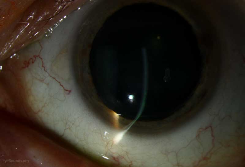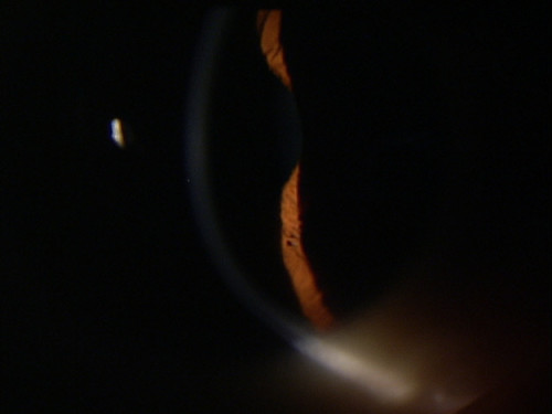

Figures 1 and 2 show the topographic images of the right and left eye, with right eye CDVA improving to 6/6 and left eye at presentation. The fellow eye was completely normal with normal topography and pachymetry. On topography (Pentacam, OCULUS Optikgeräte GmbH), the right eye showed the crab claw sign with against-the-rule astigmatism of −12.00 D at 65 degrees, and K1 (flat keratometry) and K2 (steep keratometry) were 37.90 D at 60 degrees and 51.10 D at 165 degrees, respectively. Fellow eye uncorrected distance visual acuity was 6/6 and normal on clinical examination. On examination, a crescentric arcuate irregular reflex 2.0 mm away from the limbus extending from the 4 o'clock to 8 o'clock position was noted. CASE REPORTĪ 64-year-old man presented to the clinic with blurring of vision in the right eye and was found to have an uncorrected distance visual acuity of 3/60 and a corrected distance visual acuity (CDVA) of 6/18 with a −12.00 diopter (D) cylinder axis at 65 degrees. 2 Also, only very few cases of hydrops and acute spontaneous perforation have been described in the literature. 1 Unlike keratoconus, the incidence of acute hydrops is rare, and no clear-cut modality of treatment has been described for the same. Since they are more stable within the eye, and because they mask virtually all corneal irregularity, these contact lenses offer superior optics & vision compared with any other contact lens options.ĭo you have irregularly shaped corneas, dry eyes, or a corneal disease? Talk with us about the benefits of scleral contact lenses for your vision correction! Many dozens of patients with hard-to-fit eyes or numerous corneal diseases have experienced great success by using scleral lenses here at Mt Baker Vision Clinic.Pellucid marginal degeneration (PMD) is a rare corneal ectatic disorder that is common in men in the fourth to fifth decade, which presents with bilateral and often asymmetrical peripheral corneal thinning in a crescentic manner with high against-the-rule astigmatism. You don’t need to worry about the contact lens accidentally falling out of the eye. Since the scleral lenses are bigger in size, they also stay in the eye better. Sclerals take a bit more time to insert and remove compared to most contacts, and require particular solutions to clean and fill the bowl of the lens with prior to insertion. They are also more cumbersome to use (there’s always a catch, right?).

We typically use scleral lenses that are 15.8mm in diameter, but that depends based on individual corneal sizes and conditions. How Do Scleral Lenses Compare with Other Types of Contact Lenses? From both an optics and a corneal comfort perspective, we’ve literally seen people here in Whatcom County have their lives changed due to scleral lenses.

For people with dry eye disease, incomplete lid closure, graft-host disease, or other reasons for poor corneal surface health, this can vastly improve visual clarity, as well as eye surface health and comfort. Second, the back surface of the scleral lens is fit 125-300 microns above the cornea, leaving a reservoir of lubricating saline solution continuously bathing the cornea in fluid. This allows for exceptional clarity of vision, even for people with irregular astigmatism, keratoconus, pellucid marginal degeneration, post-Lasik corneal ectasia, radial keratotomy, post-traumatic scarring, post-corneal transplant, or other forms of poor optics. First, scleral lenses create a smooth optical surface, covering over all irregularity in the cornea. There are two huge benefits to this design.


 0 kommentar(er)
